AP ELBOW IN ACUTE FLEXION
Anteroposterior projection with marked flexion for specific elbow evaluation
Special Feature of this Projection
Projection in acute flexion: This technique is used when the patient cannot fully extend the elbow due to pain, contracture, or joint limitation.
Allows evaluation of joint structures while maintaining a more comfortable position for the patient.
Exposure Factors
Slightly higher due to superposition
Low, adequate for extremity
Very low exposure: kV slightly higher (64) to compensate for structure superposition in acute flexion
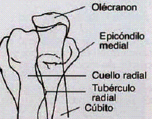
Anatomical Structures and Features
Main objective: Rule out fractures and dislocations of the elbow when full extension is not possible
Expected Superposition Feature
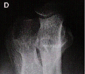
The forearm and humerus should overlap due to the acute flexion position.
An optimal exposure should show:
- Distal humerus visible through the superimposed forearm
- Olecranon visible through superimposed structures
- Joint relationship preserved despite superposition
Cassette Size
Standard size for elbow joint evaluation
Patient Positioning
Central Ray Point
Direction: Perpendicular to entry point
Location: Midpoint between medial and lateral humeral epicondyles
Consideration: Through superimposed structures in flexion
Specific Indications for this Projection
Patients with Pain
When full extension causes intense pain
Joint Contractures
Mechanical limitation of extension
Initial Evaluation
Acute trauma with movement limitation
Patient Instructions
"Remain still during the examination"
Maintain flexion position without movement during radiographic exposure
Inform technician if position causes excessive pain
Optimal Image Characteristics
Expected superposition
Forearm over distal humerus
Visible structures
Distal humerus through superposition
Olecranon visible
Identifiable process
Adequate field
Complete joint included
Common Technical Challenges
Frequent problems in AP projection with acute flexion:
- Excessive superposition hiding critical structures
- Inadequate exposure to penetrate multiple bone layers
- Position instability due to patient discomfort
- Unintentional rotation during positioning
- Exclusion of important areas due to poor centering
- Poor fracture identification due to superposition
Solution: Use slightly higher kV (64) for better penetration and ensure stable position with maximum patient comfort
Interpretation Considerations
Diagnostic Limitations
• Superposition hinders detailed evaluation
• May mask linear fractures
• Displacement evaluation limited
• Recommended to complement with other projections when possible
Technique Advantages
• Allows initial evaluation without forcing extension
• Useful in acute pain or contracture
• Can show obvious fractures or dislocations
• Less painful for patient